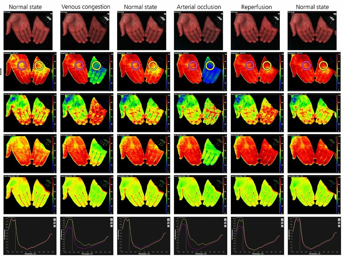
With the help of a hyperspectral camera (TIVITA®), the research project enables an objective and reproducible assessment of wounds through spatially resolved spectroscopy. In addition, the microbial load of wounds is also visualized. This is intended to extract parameters such as blood flow moisture, as well as signs of inflammation or infection of the wound in order to improve an assessment of the healing process of wounds.
Problem
Chronic non-healing wounds are an increasing problem. Physicians and nurses must rely on visual assessment to evaluate wounds because of the lack of objective descriptive methods for wounds. In order to be able to make scientifically based decisions in wound treatment, professional societies also demand that doctors be offered aid. Oxygen supply, bacterial colonization and other parameters have an influence on the healing process, but cannot be determined routinely or only with great effort and sometimes with a long time delay, for example in microbiological analysis of wound swabs.
Solution
Near-infrared spectroscopy (NIR spectroscopy) is already established in the medical environment through pulse oximetry and cerebral monitoring to measure the oxygen saturation or oxygen supply of tissue. In order to be able to objectively and reproducibly assess entire wound areas, spatially resolved spectroscopy is required. Therefore, hyperspectral camera systems suitable for clinical use have been and are being developed in the HyperWound-CAM and Bacteria-CAM projects by our collaborative partner Diaspective Vision GmbH. Model-based evaluation methods for the hyperspectral wound measurement data are being developed at Wismar University of Applied Sciences. In order to be able to determine the volume fraction of hemoglobin and its oxygen saturation in a wound segment from the remission spectra, meaningful, model-based determination methods are being developed. In addition, Monte Carlo simulations are performed to simulate the interactions of light with wound tissue. These simulations facilitate the interpretation of the spectral measurement data and enable a deeper understanding of the method. These simulations are accompanied by measurements on optical phantoms. In addition to the methods described, fluorescence excitation and detection are used to analyze microbial colonization. For this purpose, the wound area must be illuminated with UV light, while at the same time, the fluorescence is determined with spatial and spectral resolution using the hyperspectral camera. The aim is not only to determine the germ population, but also to differentiate the germs. For this purpose, preliminary investigations are to be carried out on pathogen strains, among other things, to find the optimal excitation wavelengths and the characteristic florescences. The joint projects are being carried out together with the industrial partner Diaspective Vision GmbH and the scientific partner's University of Rostock, Institute for Practical Informatics, University of Greifswald, Clinic and Polyclinic for Skin Diseases, Klinikum Karlsburg and the Competence Centre Diabetes Karlsburg.